 Aged Care Insite Australia's number one aged care news source
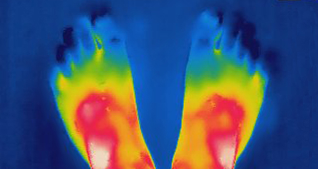
Australian researchers have used thermal imaging to assess the wound size and predict healing trajectory of diabetes-related foot ulcers – and they say it works.
Principal investigator Elif Ekinci, University of Melbourne principal research fellow and head of diabetes at Austin Health, said thermal imaging can pinpoint more accurately which wounds will heal quickly or not.
For their study, the researchers took thermal and colour images of 26 neuropathic diabetes related foot ulcers around the same time experienced podiatrists measured the wounds manually over four consecutive weeks to create a healing reference.
For healing cases, the research found the wound bed’s isothermal area was lower at two weeks compared to baseline, which corresponded with a 50 per cent reduction in area of diabetic foot ulcers at four weeks.
RMIT’s Dr Behzad Aliahmad said the study demonstrated that isothermal maps of thermal images of the wound bed from week one to week two were suitable for predicting healing trajectory.
“Thermal imaging of diabetic foot ulcers has the advantage of incorporating both area and temperature, allowing for early prediction of the healing,” Aliahmad said. “The wound monitoring by the eye of the clinician is not accurate in identifying the chronic wounds in week two.”
The study’s authors want to see thermal imaging used as an inexpensive and real-time option to identify wounds that may have delayed healing.
Do you have an idea for a story?Email [email protected]

Get the news delivered straight to your inbox
Receive the top stories in our weekly newsletter Sign up now